Orthopantomography
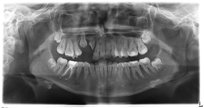
Orthopantomography (optg) is a method of x-ray examination that allows to obtain a flat one-stage image of the entire dentition system as a single functional complex.
On a correctly performed orthopantomogram, both jaws, the coronal and condyle processes of the lower jaw, the temporomandibular joint (right and left), the paranasal sinuses and part of the nasal cavity are displayed. The image of the teeth is clear, not distorted in shape. Dental cavities, periodontal cracks (especially in the premolars and molars) are clearly visible. You can distinguish a layer of enamel covering the crowns of the teeth, carious, traumatic and other defects in the tissues of the teeth.
Orthopantomography is a mandatory method of examination of the patient before orthodontic treatment.
Teleroentgenography
Telerentgenography (TRG) is the most informative method of studying the dentition, which allows you to determine the morphological changes in the facial and cerebral parts of the skull, to justify the diagnosis and treatment plan.
Analysis of teleroentgenogram allows to solve the following tasks:
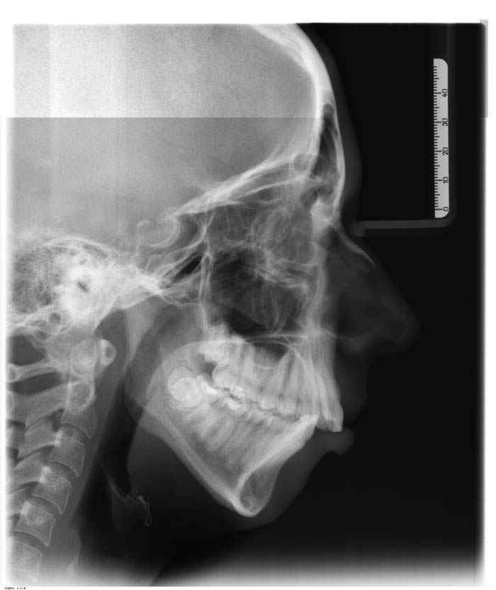
- to determine the facial profile of the patient;
- determine the size of the jaws and their apical bases;
- determine the position of the jaws relative to the base of the skull;
- determine the relative position of the jaws;
- determine the type of growth of the facial skeleton;
- to assess the position of the teeth and the development of alveolar processes;
- to conduct a differential diagnosis of clinical varieties of malocclusion;
- make a final diagnosis;
- justify the plan of treatment of the patient.
 
Radiography of TMJ
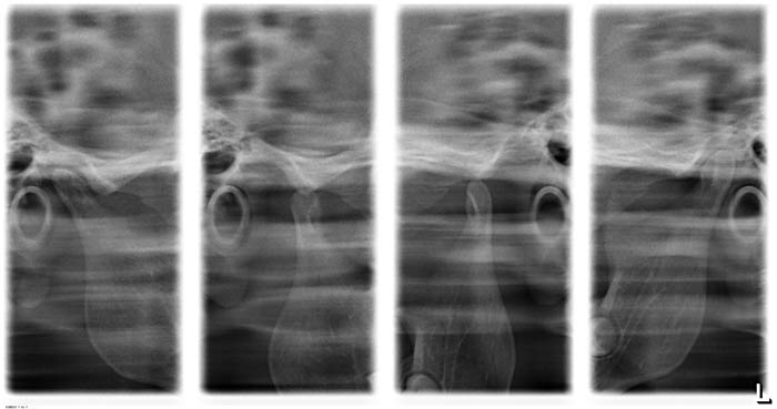 The method allows the state of the closed dentition and the maximum opening of the mouth. It is an Express method for the diagnosis of TMJ. For a detailed examination of the shape, position, condition of the heads of the lower jaw, articular fossa, disc and ligamentous apparatus of the joint, a computer tomography is necessary to visualize the position of the heads of the temporomandibular joint in or magnetic resonance imaging.
 
Cone-beam computed tomography

Cone beam computed tomography (CBCT) is a modern x-ray examination method, which is a kind of 3D computed tomography. The method has a very high informative and significantly expands diagnostic capabilities in such areas of medicine as dentistry, otorhinolaryngology and maxillofacial surgery.
With the help of the CBCT method, unlike the optg and TRG, the visualization of the area of interest in the volume takes place. This allows to obtain a three-dimensional model, to build any cross-section, to carry out very accurate measurements. Other advantages of CBCT are lower radiation load and the absence of overlays of anatomical structures and distortions of their size.
 
Electromyography
Electromyography (EMG) is an objective method of studying the neuro-muscular system by registering the electrical potentials of the masticatory muscles, which allows to assess the functional state of the dentoalveolar system. The electrical activity of the masticatory muscles is recorded simultaneously on both sides. Surface electrodes are used to remove biopotentials. The electrodes are fixed in the area of the motor points of the masticatory and temporal muscles (areas of the greatest muscle tension, which are determined by palpation). Recording of EMG is performed at rest and when performing functional tests (with jaw compression in the right, left, in the usual occlusion, opening/closing of the mouth, movements of the lower jaw forward/backward. Normally, there is a coordinated function of muscle synergists and antagonists, a clear rhythmic change of phases of bioelectric activity and bioelectric rest. Chewing and temporal muscles during chewing show synchronous activity. Interpretation of the obtained EMG data is possible in a comprehensive study of the dentoalveolar system, since the same changes in the EMG pattern occur in various pathological conditions (loss of teeth, malocclusion, decrease in occlusive height, bruxism).


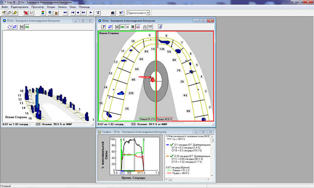
Occlusion analysis using T-scan
To determine the occlusive effort, the patient is invited to bite the individual thin sensor. On the computer monitor there is an image in the form of a video showing all the movements of the teeth from the first to multiple contact. The obtained data are displayed on two-dimensional and three-dimensional active diagrams, according to which the specialist can not only quickly and accurately see the problem points (premature contact, too strong contact), but also clearly show the patient the cause of the pathology.
 ¬†¬†
¬†¬†
¬†¬†